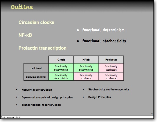
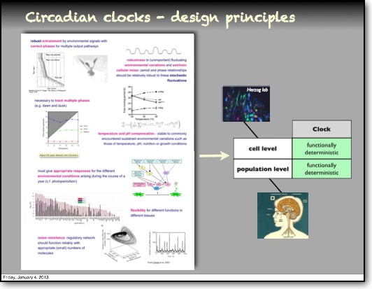
The NF-κB System
Nuclear Factor kappa B (NF-κB)
NF-κB controls inflammation
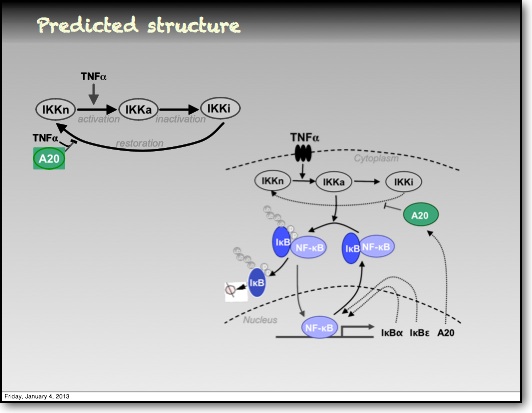
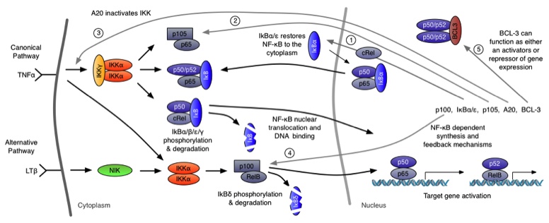
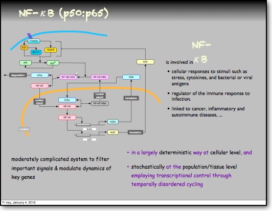
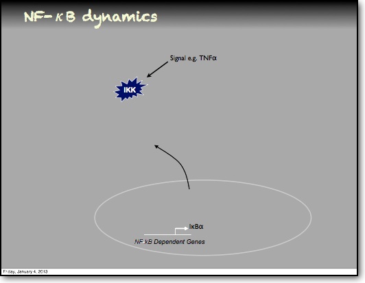
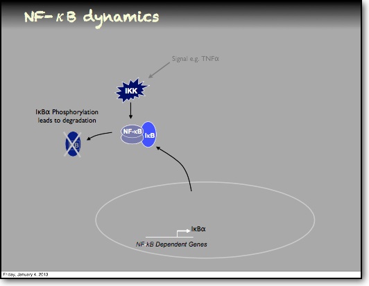
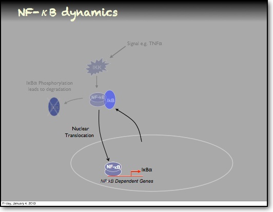
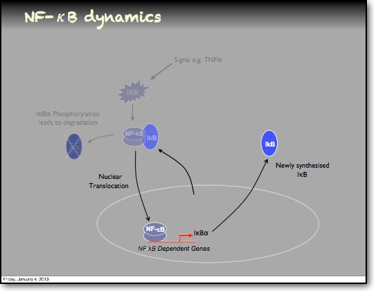
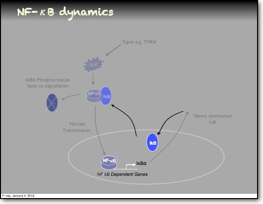
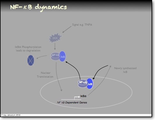
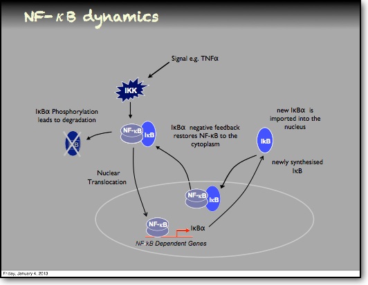
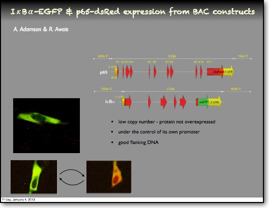
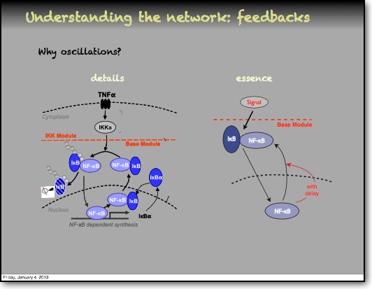
Discovered 25 years ago, the NF-κB transcription factor controls inflammation and in different contexts has varying effects on cell death and cell division. It involves a family of proteins and NF-κB binding sites are found in the promoter regions of around 300 genes, including cytokines (e.g. TNFα, LTβ, IL-1 and GM-CSF). NF-κB is activated by various stress stimuli, including inflammatory cytokines such as TNFα and IL-1β. NF-κB signalling involves phosphorylation of IκB proteins by the IKK kinase complex. This allows the translocation of an NF-κB dimer (typically RelA:p50) into the nucleus. Target genes include IκBα and IκBε, which are therefore feedback inhibitors of NF-κB signalling. Their delayed expression gives the system the characteristics of a delayed feedback oscillator.Oscillatory dynamics of NF-κB visualised
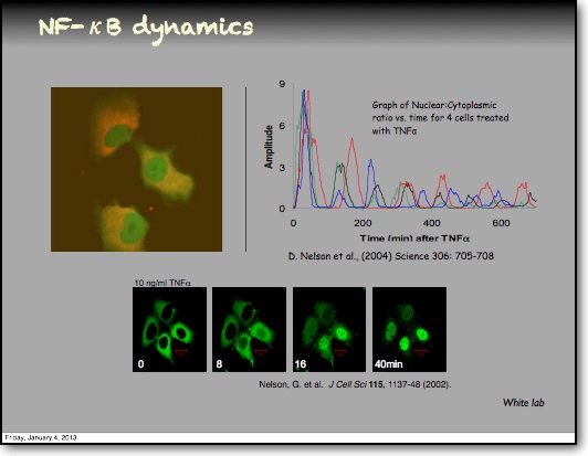
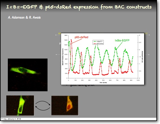
The White lab used NF-κB fluorescent fusion proteins to visualise and quantify the oscillatory dynamics of NF-κB signalling by single cell timelapse imaging. As a result of these and other subsequent studies, NF-κB has become perhaps the best-studied protein-based oscillatory signalling system. A key feature is the robust 100 min oscillations observed in cells (Fig.). This period has appeared to be the most stable feature of NF-κB signalling in response to varying levels of TNFα stimulation. We showed however that forcing the oscillations with pulsatile TNFα stimulation could give rise to a range of frequencies of NF-κB translocation, which depended on the stimulation frequency.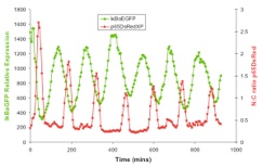
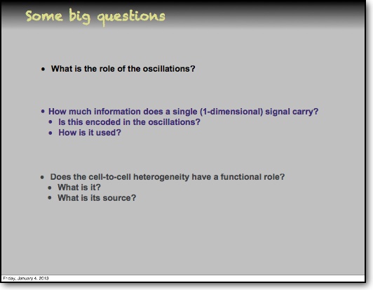
Feedback and the timing of oscillations
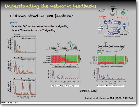
In addition to the IκBα and IκBε there are other feedbacks from a set of genes for proteins that act upstream from IKK, the most studied being the protein A20. The timing of oscillations in response to TNFα is remarkably consistent between cells. However, slight variation in the timing between peaks of translocation causes cells to quickly fall out of synchronisation with each other2. It has been proposed that transcriptional noise in negative feedback genes (there being only 2 gene copies of each) may explain this lack of synchronicity between the cells. We used a combined modelling and experimental strategy to suggest that the transcriptional activation of the IκBε feedback loop is delayed relative to the IκBα feedback loop by around 40 min. Analysis of the mathematical model suggested that this timing lay at a theoretical optimum for the generation of heterogeneity between cells and thus might be an evolutionarily selected phenotype. Such heterogeneity might minimise the overall fluctuations in cytokine levels in a tissue and therefore be important in accurate control of the tissue-level inflammation. This raises the interesting idea that overall tissue level inflammation may be in part regulated by the degree of heterogeneity between individual cells. This potentially represents a new phenotype that might be a possible drug target to manipulate the balance between hypo- and hyper-inflammation.Some Recent Papers
2010 Lecture at BBSRC Grant Holders Meeting in Edinburgh
Your Headline Here
Your description goes here.
Your Headline Here
Your description goes here.
Your Headline Here
Your description goes here.
Your Headline Here
Your description goes here.
Your Headline Here
Your description goes here.
Your Headline Here
Your description goes here.
Your Headline Here
Your description goes here.
Your Headline Here
Your description goes here.
Your Headline Here
Your description goes here.
Your Headline Here
Your description goes here.
Your Headline Here
Your description goes here.
Your Headline Here
Your description goes here.
Your Headline Here
Your description goes here.
Your Headline Here
Your description goes here.
Your Headline Here
Your description goes here.
Your Headline Here
Your description goes here.
Your Headline Here
Your description goes here.
Your Headline Here
Your description goes here.
Your Headline Here
Your description goes here.